Biological Sciences 330, Smith College | Research in Cellular Neurophysiology
Swimmeret Central Pattern Generator
Revised: February 19, 2019
|
View and listen to recordings of the swimmeret pattern in response to different concentrations of carbachol. |
|
Experimental methods.
View the video: Central Pattern Generator for Crayfish Swimmerets, which shows the procedure for isolating a crayfish nerve cord and recording from the first roots.
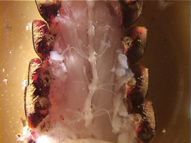
Recording from an isolated crayfish nerve cord with pin electrodes enables us to detect the patterned activity produced in the first roots during "fictive locomotion" of the swimmerets generated by central pattern generator circuits in the ganglia. The swimmeret muscles receive axons from motoneurons in root 1 of the second through the fifth abdominal ganglia. Recordings can be made from a first root of a semi-intact preparation using suction electrodes, but you will get better control over drug concentrations if you record from an isolated nerve cord using the pin and vaseline electrode method described below.
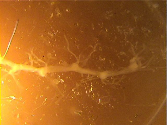
To prepare an isolated nerve cord, follow the procedures outlined in an earlier lab to expose the abdominal nerve cord, and then continue with the dissection shown in the video for this lab. In cutting all the roots, be especially careful to cut the first roots (N1) as far from each ganglion as possible, while you cut the other roots short. This will avoid confusion about which roots are N1s after the cord is isolated.
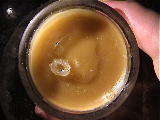
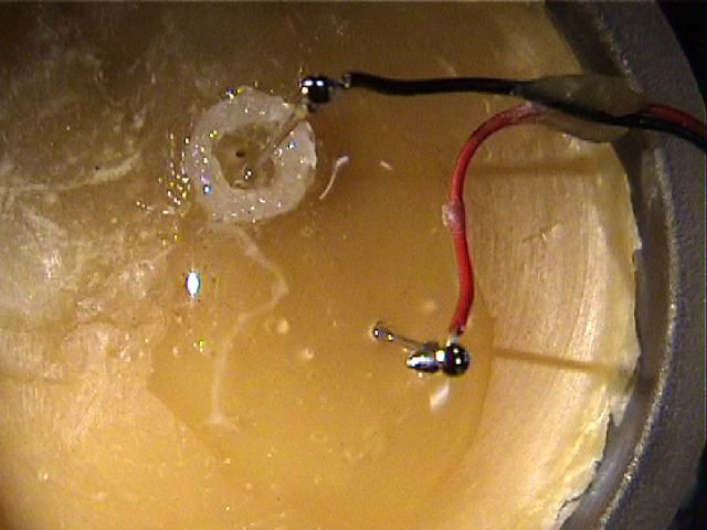
On a small dry dissecting dish, make a ring of vaseline that is tall enough to hold back the saline that you will later place in the dish. Move the dissected nerve cord into the dry dish, and drape the branch or branches of a first root through the vaseline so the cut end of the root lies inside the circle. Add a drop or two of saline inside the ring, and create a pool of saline around the main part of the nerve cord. You may also wish to pin out each end of the nerve cord using fine minutenadeln ("tiny needles").
Place pin electrodes in the main pool of saline and in the center of the vaseline ring. The pin and vaseline-ring combination functions like a suction electrode. The vaseline is like the tight-fitting tip of a suction electrode, separating the saline inside the ring from the saline outside. As local circuit currents from action potentials in the axons move through the vaseline region, the wires inside and outside the ring detect a difference in potential. To get good recordings of spikes, it is important that there not be a leak through the vaseline.
To apply drugs, gently remove the main pool of saline around the nerve cord using a transfer pipette. Add some of the new drug solution as a wash, remove it, and add the new drug solution again. The drug will diffuse into the ganglia and reach the synaptic regions in the neuropil. Note that drugs are applied to the main pool surrounding the nerve cord, so that they can diffuse into the ganglia where the synapses are located. Drugs are not placed in the recording wells, where they would reach only the cut ends of axons.
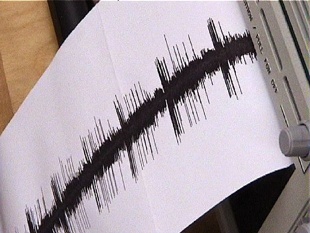
Monitor neural activity on your oscilloscope screen, your audio monitor, your computer (using PowerLab), and directly on your chart recorder. Rhythmic CPG bursts will be easy to hear, and you will see the bursting pattern easily on the computer screen (at slow scrolling speeds) or on your chart recorder. PowerLab will be the most versatile, since you can later zoom in on details of the activity, integrate the records or analyze spike rates, and create materials for your presentation.
Some helpful hints:
- The pin electrodes have very delicate wires,
so treat them gently. If a wire or pin breaks off, let
the instructor know. They are easy to fix.
- We have fine dissecting scissors that also
need to be treated very gently. No stabbing them into
your dissecting dish in a moment of frustration! Be sure
to wash and try them carefully at the end of each
afternoon.
- Drug solutions are always made up and diluted
in crayfish saline, not distilled water. Again,
note that drugs are applied to the main pool
surrounding the nerve cord, not in the recording
wells.
- If you plan to combine two drugs, make up your stock solutions of
each individual drug at twice the highest
concentration you plan to use. To test one drug at its
highest planned concentration, dilute your stock
solution 1:1 with saline. To combine the drugs, mix the
two stock solutions in suitable proportions. For example,
mixing the two stock solutions 1:1 will create a new
solution in which each drug's stock concentration has
been diluted in half.
- Work with small volumes. You only need a few
ml to create a pool around the nerve cord, and some drugs
are expensive. The only reason to make more than 5 or 10
ml of a solution is if you need to make a big dilution
(eg, 1:100) of a primary stock solution. See about
joining forces with other groups who are using the same
drugs that you are.
- Avoid test-tube build-up. Resist the
temptation to make a large number of dilutions in advance
-- you may end up wasting many of them. Instead, make
dilutions when you need them (it's a very quick process).
Usually, you can make serial dilutions, which also helps
avoid large volumes. We have calibrated centrifuge tubes
(15 ml and 50 ml) and calibrated transfer pipettes (1 ml)
for making dilutions.
- Label your solutions AND keep them together in a beaker labeled with your group's names and lab day. Label tape and markers are available.
Links
Supplement: Anatomy of the Crayfish Nervous System.
Background information about the crayfish swimmeret system.
Appendix: Capturing Oscilloscope Screenshots
Appendix: Using EasyGraf Chart Recorders.
© 2003 - 2016, 2019 by Richard F. Olivo. Permission is granted to non-profit educational institutions to reproduce or adapt this Web page for internal use provided that the original source and copyright are acknowledged.