Biological Sciences 330, Smith College | Research in Cellular Neurophysiology
Anatomy of the Crayfish Nervous System
Classic drawings
General anatomy of the crayfish nervous system. Source: Cattaert and LeRay (2001) Adaptive motor control in crayfish. Progress in Neurobiology 63: 199-240.
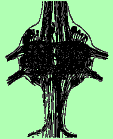
Crayfish brain (Retzius, 1890) Source: Nassel and Elofsson, in Gupta, 1987, Arthropod Brain (Wiley).
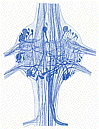
Diagram of lobes in crayfish brain (Helm, 1928) Source: Nassel and Elofsson, in Gupta, 1987, Arthropod Brain (Wiley).
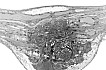
Third abdominal ganglion stained with methylene blue (Retzius, 1890) Source: Bullock and Horridge, 1965, Structure and Function in the Nervous System of Invertebrates (Freeman).
Videos of serial sections through an abdominal ganglionby Brian Mulloney and Wendy Hall, |
 |
|
|
|
|
|
|
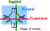
Transverse sections (cut perpendicular to the long axis of the nerve cord). The sections move from the connective at the anterior of the ganglion to the connective at the posterior end. |
Sagittal sections (vertical sections cut parallel to the long axis of the nerve cord). The sections move from the roots on one side to the roots on the other side. |
Frontal sections (cut parallel to the ventral surface of the abdomen). The sections move from the ganglion's ventral surface to its dorsal surface. |

Reference: Mulloney, B., T. Naranzogt and W.M. Hall (2003) Architectonics of crayfish ganglia. Microsc. Res. Tech. 60: 253-265.
We thank Professor Brian Mulloney, University of California at Davis, for permission to include reformatted versions of his videos and figures from his papers.
Stained swimmeret motoneurons
Several cobalt-stained motoneurons in a first root, showing the position of their cell bodies near the base of the nerve and the branching of their processes in the lateral neuropil. Source: Sherff, C.M., B. Mulloney (1997) Passive properties of swimmeret motor neurons. J. Neurophysiol. 78:92-102.
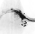
Heavily stained cobalt
backfills of the first root on the left and right
sides of a third abdominal ganglion.
Source: Mulloney, B. and W.M. Hall (2000)
Functional organization of crayfish abdominal ganglia: III.
Swimmeret motor neurons. J. Comp. Neurol. 419:
233-243, Fig. 2.
A cobalt backfill of a first root in which most of the axons in the nerve filled well. (Mulloney & Hall 2000, Fig. 3)
Another backfill of a first root in which only a few axons filled. (Mulloney & Hall 2000, Fig. 3)
Backfill of the posterior branch of a first root, showing the position of power stroke motoneurons in the ganglion. (Mulloney & Hall 2000, Fig. 4)
Backfill of the anterior branch of a first root, showing the position of return stroke motoneurons in the ganglion. (Mulloney & Hall 2000, Fig. 4)
© 2003 by Richard F. Olivo. Permission is granted to non-profit educational institutions to reproduce or adapt this Web page for internal use provided that the original source and copyright are acknowledged.