What cell types help to establish connections between the two sides of the brain?
Bilaterally symmetrical organisms require proper communication between the left and right sides of the body. This communication is established through neuronal structures called commissures. The most common examples of commissures in humans are the optic chiasm and corpus callosum, which are both vitally important for coordination and sensory processing between the two sides of the brain. Understanding how neurons navigate the embryonic brain to build commissures can help us to identify the key parameters that may be needed to grow and guide such wiring following neurodegenerative disorders and diseases.
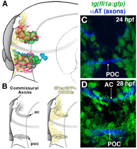
Zebrafish also have an optic chiasm and similar brain commissures, but also provide a tractable and simple system to get at the molecular mechanisms controlling this process. We have a long history of studying the role that astroglial cells play in forming a permissive substrate for commissure formation, as well as defining the Slit-Robo signaling system in guiding neuronal axon pathfinding across the midline all in the zebrafish forebrain. Watch a movie of axons forming a commissure in the zebrafish forebrain.
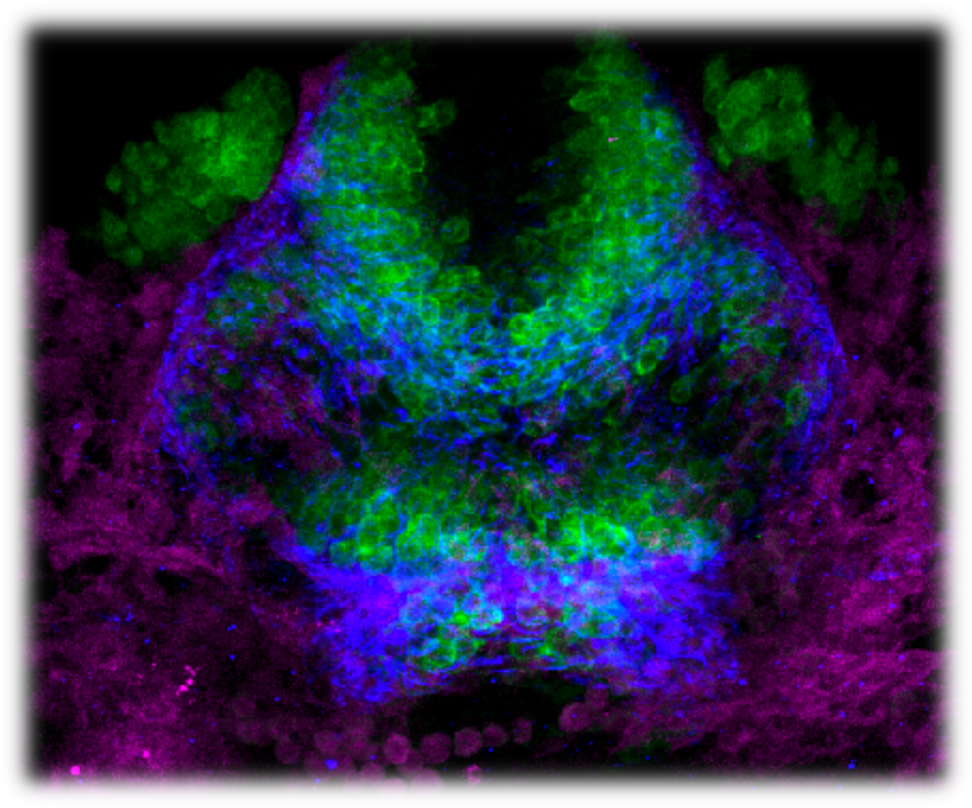
 More recently, we completed a comprehensive characterization of the varied cell types that interact with pathfinding commissural axons during forebrain development. We discovered several populations of early neuronal, glial, and progenitor cells that directly interact with the forming commissure. Surprisingly, we discovered that a transient population of migrating stem cells, known as cranial neural crest cells, prefigured and overlapped with the developing commissure.
We are currently characterizing what these migrating stem cells are doing to pattern the forebrain. Do they provide signals to guide commissural axons? Although cranial neural crest cells are well-known to give rise to cranial facial structures (bones of the face in humans and fish alike), could these cells be migrating into the forebrain and directly contributing to neurogenesis?
Although first born from neural ectoderm, neural crest cells have been thought to remain peripheral to the CNS and contribute only to peripheral structures. Demonstrating that neural crest cells play a direct role in patterning the brain and contributing to neurogenesis would be a paradigm shift in our knowledge of the ontogeny of these amazing cells. See a video of cranial neural crest cells migrate toward the forebrain.
This work has been funded by the National Science Foundation.