|
Clinozoisite
|
|
Property
|
Value
|
Comments
|
| Formula |
Ca2Al3O(SiO4)(Si2O7)(OH)
|
Forms a solid solution series with epidote by substituting Al+3 for Fe+3 |
| Crystal System |
monoclinic; 2/m |
|
| Crystal Habit |
rounded grains, columnar or radial aggregates |
Elongate parallel to the b axis |
| Cleavage |
{001} perfect |
Often controls fragment orientation. Can be difficult to observe due to small grain size. |
| Hardness |
7 |
| Specific Gravity |
3.3 - 3.4 |
| Color/Pleochroism |
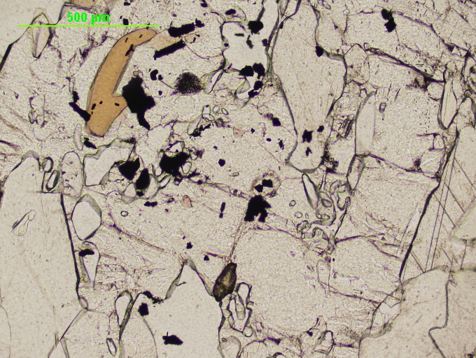
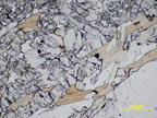
pale green to gray in color, transparent to translucent; not pleochroic.
|
| Optic Sign |
Biaxial (+) |
|
| Birefringence |
0.006-0.0011 |
|
| 2V |
14° - 90° |
Often > 65 |
Refractive Index
|
alpha = 1.706 – 1.724
beta = 1.708 – 1.729
gamma = 1.712 – 1.735
|
Refractive indices and birefringence increase with iron content. |
| Extinction |
Extinction angle to trace of cleavage usually between 0° & 25°. |
Sometimes the extinction angle can be as large as 60°. |
| Distinguishing Features |
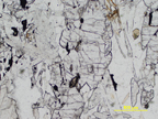
Distinguishable from epidote in thin section by optic sign, lower birefringence, and absence of green color. Clinozoisite presents maximum first order colors under cross-polarized light and is mostly colorless in plain polarized light.
|
| Occurrence |
Commonly present in subduction zones and along contacts of Ca-rich and Al-rich rocks. Where iron is present, epidote will be formed instead. Clinozoisite and epidote are common accessory minerals in regional and contact metamorphic rocks. It can be present in quartzite, slate, chlorite schist, mica schist, gneiss, amphibole, marble, skarn deposits, hornfels, phyllite, and calc-silicate.
|
| References |
Nesse, William D., "Introduction to Optical Mineralogy." Oxford University Press: New York, 2000. pp. 295-296
"Clinozoisite: Clinozoisite Mineral Information and Data." Mineralogy Database - Mineral Collecting, Localities, Mineral Photos and Data. Web. 27 Jan. 2011. <http://www.mindat.org/min-1087.html>. |
| Editors |
Abby D'Ambrosia ('07), Lilly Dalton ('11) |