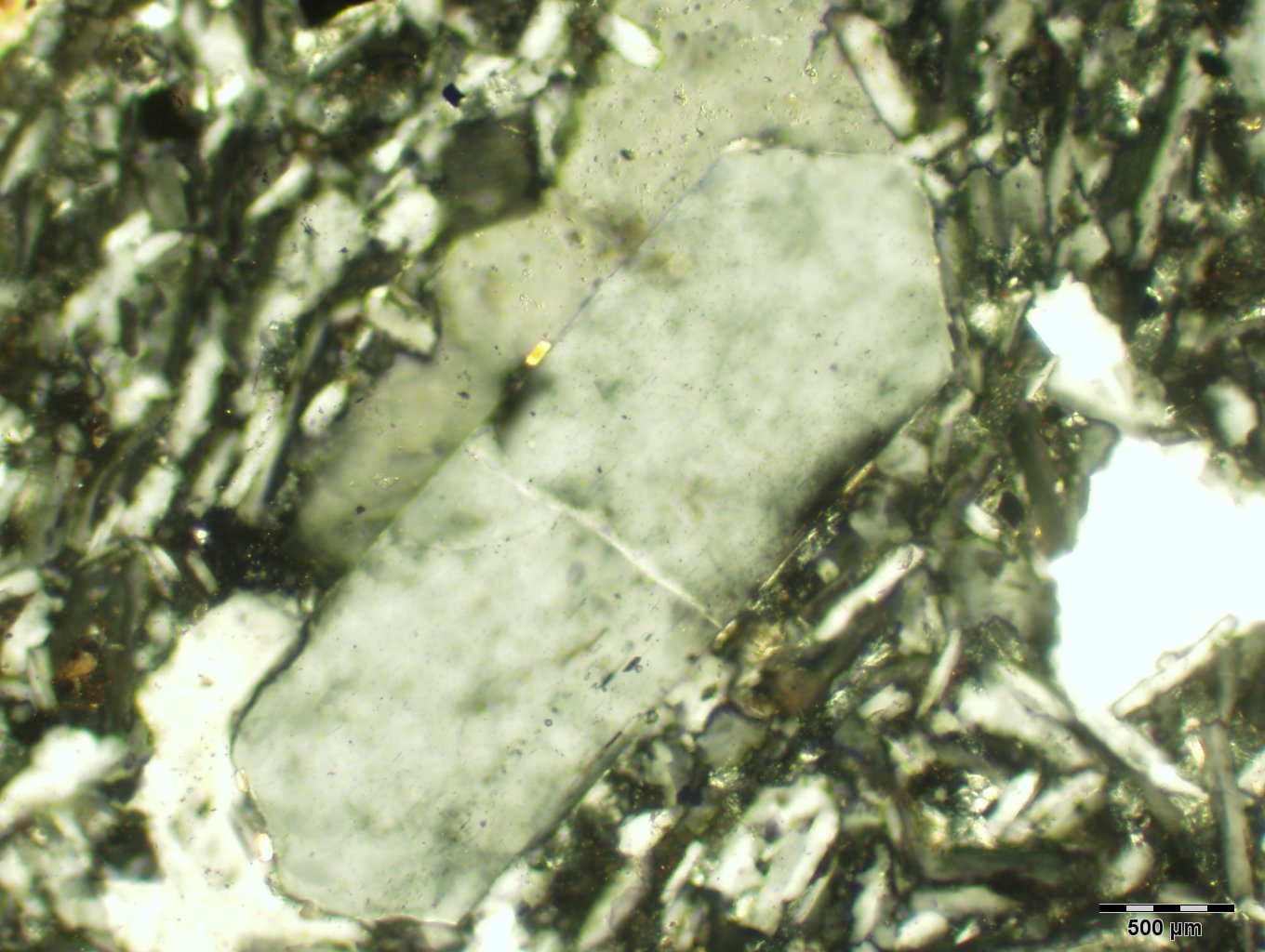
Sanidine |
|
Property |
Value |
Comments |
| Formula |
KAlSi3O8 |
High temperature (above 700°) sister to orthoclase and
microcline. Solid solution of sodium (albite) possible, but not common.
The difference between sanidine, orthoclase, and microcline is its random
order of placement of Al within bonding sites. Microcline and Orthoclase
have ordered placement of Al. |
| Crystal System |
Monoclinic |
Another way to tell the difference between sanidine, orthoclase,
and microcline is that sanidine and orthclase are monoclinic and microline
is triclinic. |
| Crystal Habit |
prismatic, tabular |
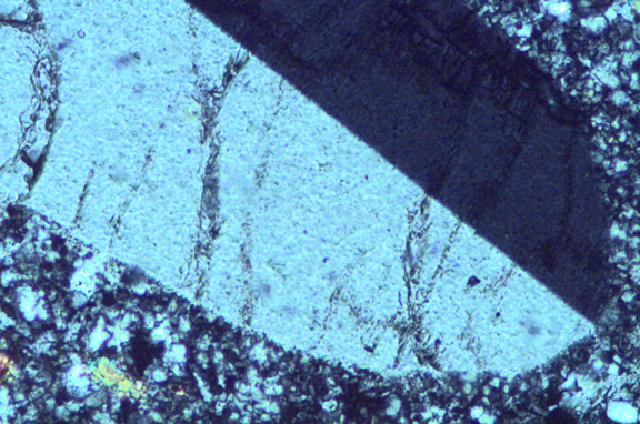
Zoning is common, expressed in varying birefringence and
extinction angles. |
| Cleavage |
(001) perfect; (010) good |
|
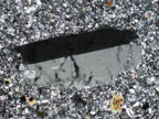
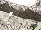
| Twinning |
Carlsbad twinning with (010) composition plane dividing crystal
into two segments. |
Manebach and Baveno twins also possible. |
| Color/Pleochroism |
Colorless in thin section. |
|
| Optic Sign |
Biaxial (-) |
|
| 2V |
0-40° |
Lower temperatures produce an increase of Al and Si in tetrahedral
sites, increasing 2V values. Na substitution can also increases the
2V. |
| Optic Orientation |
X^a = +5 to +9 °
Y^c = +21 to +17°
Z = b
O.A.P. normal to (010) |
High temperature sanidine has a differently oriented optic
plane:
X^a = +5°
Y = b
Z^c = +21°
O.A.P. parallel to (010) |
Refractive Indices
alpha =
beta =
gamma =
delta = |
1.514 - 1.526
1.518 - 1.530
1.521 - 1.533
0.005 - |
|
| Birefringence |
0.005 to 0.008 |
No higher than first order white |
| Elongation |
somewhat elongate parallel to the a axis |
|
| Extinction |
Can be parallel |
|
| Dispersion |
r > v |
|
| Distinguishing Features |
Generally distinguished from orthoclase by smaller
2V. High sanidine indicated by orientaion of optic plane. |
| Occurrence |
Formed in high temperature volcanic and hypabyssal igneous
rocks. Can also be present in high tempereature contact metamorphic
rocks. |
| Editors |
Amanda Trotter (AC), Kristin Abel (02), Robyn Bluestein
(03), Lucy Eckert (05), Roxanne Renendo (09), Alyssa Pascuzzo (15) |