|
Sillimanite
|
|
Property
|
Value
|
Comments
|
| Formula |
Al2SiO5 |
Most sillimanite is relatively pure, although minor amounts of Fe3+, Cr3+, Ti4+ may be present. |
| Crystal System |
orthorhombic |
a=7.488, b=7.681, c=5.777. |
| Crystal Habit |
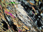
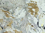
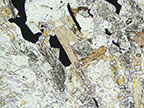
Slender prismatic or fine fibrous crystals. |
Masses of fine fibrous crystals, known as fibrolites, may form swirled or matted aggregates. Prismatic crystals crystals may appear rectangular or diamond shaped in cross section with diagonal cleavage lines. |
| Cleavage |
Perfect on (010)
|
Uneven fracture. Brittle. Fibrous aggregates may be tough. |
| Color/Pleochroism |
Colorless or white, less commonly yellow, brown or blue. |
Vitreous luster and white streak. |
| Optic Sign |
Biaxial (+) |
|
| 2V |
20-30 |
|
| Optic Orientation |
X=a
Y=b
Z=c
|
- Optic axis dispersion strong, r > v.
- Fragments on (010) cleavage yield flash figures. |
Refractive Indices
alpha =
beta =
gamma =
delta = |
1.653-1.661
1.657-1.662
1.672-1.683
0.018-0.022 |
|
| Max Birefringence |
0.018-0.022 |
Indices of refraction show little variation. Interference colors in thin section, are up to lower second order. Fine fibrolite fibers show lower inerference colors because they are hinner than a standard thin section. |
| Elongation |
length slow |
|
| Extinction |
parallel |
|
| Distinguishing Features |
High relief, moderate birefringence, parallel extiction, and slender prismatic or fibrous crystal habit. Distinctive diamond-shaped cross sections with cleavage for prismatic grains. Fibrous mats commonly replace biotite. |
| Occurrence |
Sillimanite occurs as a constituent of high-temperature metamorphosed rocks containing clay. In contact metamorphosed rocks it may occur in silimanite-cordierite gneisses or sillimanite-biotite horfels. In silica poor rocks, it may be associated with corundum. |
| Editors |
Mai Kobayashi ('06), Lyn Watts ('17) |